
A twisted ankle that never quite feels right, or a knee that catches and swells after activity, can sometimes point to more than a simple sprain or strain. Osteochondral defects – injuries that involve both the smooth cartilage surface of a joint and the underlying bone – are a recognized cause of persistent joint pain, particularly in the knee and ankle. Because these injuries affect tissue with limited natural healing capacity, understanding the available options matters for anyone dealing with ongoing joint symptoms after an injury.
This article reviews what osteochondral defects are, how they develop, and the staged conventional treatments typically used, from conservative management through surgical cartilage restoration. It also looks at how regenerative approaches, including mesenchymal stem cells (MSCs) and platelet-rich plasma (PRP), are being studied as complementary options for osteochondral defect repair, what current research shows, and what realistic expectations look like for anyone exploring osteochondral defect treatment.
What Are Osteochondral Defects?
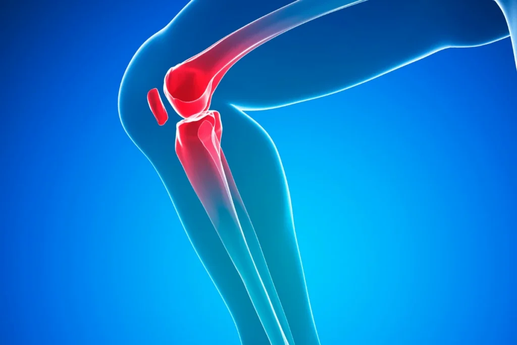
Joints like the knee and ankle are lined with articular cartilage – a smooth, low-friction tissue that allows bones to glide against one another – sitting atop a layer of subchondral bone that supports it. An osteochondral defect occurs when both this cartilage layer and the bone beneath it are damaged, creating a lesion that goes deeper than a typical cartilage injury. Because mature cartilage has very limited blood supply and a low capacity for self-repair, these combined cartilage-and-bone injuries often do not heal well on their own.
The knee (particularly the femoral condyles) and the ankle (specifically the talus, where these injuries are often called osteochondral lesions of the talus, or OLT) are the most common sites. Causes generally fall into a few categories: acute trauma such as an ankle sprain, twisting injury, or fall; repetitive mechanical stress from high-impact sports; and osteochondritis dissecans, a condition in which a piece of cartilage and bone can partially or fully separate from the surrounding joint surface, often in younger, active patients. Symptoms typically include localized joint pain that worsens with activity, swelling, stiffness, a sense of catching or locking, and in some cases a feeling of instability, though small defects can also be asymptomatic and discovered incidentally.
Conventional Treatment Options
Treatment for osteochondral defects is generally staged according to lesion size, location, symptom severity, and patient activity level.
Conservative management is typically the first step for smaller, stable, or minimally symptomatic lesions. This may include activity modification, protected weight-bearing, physical therapy to support surrounding muscle strength and joint mechanics, bracing, and anti-inflammatory strategies. A structured trial of nonsurgical care is often attempted before moving to invasive options, especially in less severe cases.
Arthroscopic debridement may be used to clean up loose or damaged cartilage fragments and address associated joint irritation, sometimes as a first surgical step or alongside other techniques.
Microfracture is a bone marrow stimulation technique in which small perforations are made in the subchondral bone to release marrow elements, including bone marrow-derived stem cells, into the defect, prompting formation of fibrocartilage repair tissue. It is generally reserved for smaller lesions, as research has found that outcomes may be less durable for larger or more complex defects compared with restorative procedures (Angele et al., Cartilage, 2022).
Osteochondral autograft and allograft transplantation (OATS/OCA) involves transferring a plug of healthy cartilage and underlying bone, either from the patient’s own body (autograft) or from a donor (allograft), into the defect site. This restorative approach is often considered for larger lesions or when marrow stimulation techniques have failed.
Autologous chondrocyte implantation (ACI) and matrix-induced ACI (MACI) are two-stage procedures in which a patient’s own cartilage cells are harvested, expanded in a lab, and then reimplanted, often on a supportive scaffold. These techniques are generally used for larger, full-thickness defects and are associated with improved knee function and pain scores across multiple studies (Nassar et al., Knee Surgery, Sports Traumatology, Arthroscopy, 2025). For a closer look at nonsurgical pathways patients sometimes explore before considering these procedures, see this overview of nonsurgical knee cartilage injury treatment options.
How Regenerative Medicine May Support Cartilage Repair
Alongside conventional surgical and nonsurgical care, regenerative medicine approaches are increasingly being studied as complementary options for osteochondral defect repair. Mesenchymal stem cells (MSCs), sourced from bone marrow or adipose tissue, are of particular interest because they have chondrogenic potential – the capacity to differentiate toward cartilage-forming cell lineages under the right conditions – and appear to exert paracrine anti-inflammatory signaling, meaning they may release factors that modulate local inflammation and support a more favorable healing environment, rather than simply replacing tissue directly.
Platelet-rich plasma (PRP), a concentrate of a patient’s own platelets and growth factors, is another regenerative option being explored for osteochondral defects, including osteochondral lesions of the talus. PRP is thought to support the joint environment by contributing growth factors involved in tissue repair and modulating local inflammatory activity. Bone marrow aspirate concentrate (BMAC), which contains a mixture of marrow-derived cells including MSCs, has similarly been studied as an adjunct to marrow stimulation and other cartilage procedures.
It’s important to be clear that these regenerative approaches are considered complementary and emerging rather than first-line treatments for osteochondral defects. Evidence supporting their use for cartilage repair is still developing, and current research consists largely of small clinical trials, case series, and mechanistic studies rather than large, definitive randomized trials. They are not a substitute for an accurate diagnosis and an individualized treatment plan developed with an orthopedic or regenerative medicine provider.


The Evidence: What Research Shows
Research on osteochondral defect treatment spans conventional cartilage restoration and emerging regenerative options.
A systematic review of Level 1 studies with at least five years of follow-up found that restorative techniques such as osteochondral autograft transplantation were associated with better long-term outcomes and lower failure rates compared with microfracture for full-thickness knee cartilage defects (Angele et al., Cartilage, 2022). A more recent systematic review and meta-analysis similarly found that ACI, MACI, OATS, and osteochondral allograft transplantation were each associated with meaningful improvements in knee function and pain, with outcomes varying based on defect size and patient characteristics (Nassar et al., Knee Surgery, Sports Traumatology, Arthroscopy, 2025).
For osteochondral lesions of the talus specifically, a narrative review of clinical treatment advances noted that microfracture continues to be favored for smaller lesions, while replacement or biologic-augmented strategies are increasingly used for larger or cystic defects (Liao et al., Orthopedic Surgery, 2025). A separate systematic review of clinical trials using PRP for osteochondral lesions of the talus found that PRP was generally well tolerated and associated with symptomatic improvement in several small studies, though the authors noted that study quality and sample sizes limited firm conclusions (Yausep et al., Journal of Orthopaedics, 2020).
On the stem cell side, a clinical trial using allogeneic umbilical cord blood-derived MSCs combined with a hyaluronate hydrogel for cartilage defects reported safety and encouraging structural outcomes over seven years of extended follow-up (Park et al., Stem Cells Translational Medicine, 2017). A broader review of clinical trials examining MSC therapies for cartilage regeneration in osteoarthritic joints concluded that while intra-articular MSC applications appear generally safe, current evidence suggests they may be insufficient on their own to achieve full regeneration of damaged articular cartilage, underscoring the need for continued research (Carneiro et al., International Journal of Molecular Sciences, 2023). Readers interested in how these regenerative concepts are being applied to ankle injuries specifically may find this discussion of stem cell therapy for ankle injuries useful background.
Taken together, this body of research indicates that conventional restorative surgery has the strongest and longest track record for osteochondral defect treatment, while regenerative options remain a promising but still-developing area of study.
Safety Considerations and Realistic Expectations
Accurate diagnosis is the essential first step before considering any osteochondral defect treatment. This typically involves a clinical history and physical exam, imaging such as X-ray and MRI to characterize the size, depth, and location of the lesion, and in some cases diagnostic arthroscopy to directly visualize the joint surface. Lesion size, stability, and location strongly influence which treatment pathway is appropriate.
It’s also important to understand the regulatory status of regenerative options. Mesenchymal stem cell therapy and PRP for cartilage and osteochondral defect repair are not FDA-approved treatments for this indication; they are used and studied as part of ongoing clinical research and individualized care rather than as established, approved therapies. This does not mean they lack value, but it does mean expectations should be grounded in the current evidence rather than assumptions of guaranteed results.
Anyone experiencing persistent joint pain, swelling, or mechanical symptoms such as catching or locking should consult a qualified orthopedic or regenerative medicine provider for evaluation. A provider can help weigh conservative care, surgical restoration, and regenerative options based on the specific characteristics of the defect and the individual’s activity goals and overall health.
Frequently Asked Questions
What is an osteochondral defect?
An osteochondral defect is an injury that involves both the articular cartilage covering a joint surface and the underlying subchondral bone. It differs from a purely cartilage-only injury because the damage extends into the bone layer beneath the cartilage, which can complicate healing.
Where do osteochondral defects most commonly occur?
The knee (especially the femoral condyles) and the ankle (specifically the talus) are the most frequently affected sites. Ankle lesions are often referred to as osteochondral lesions of the talus, or OLT.
What causes osteochondral defects?
Common causes include acute trauma such as ankle sprains or twisting injuries, repetitive mechanical stress from high-impact activity, and osteochondritis dissecans, a condition where a fragment of cartilage and bone can loosen from the joint surface.
What is the first-line treatment for a smaller osteochondral defect?
Smaller, stable lesions are often managed initially with conservative care, including activity modification, physical therapy, and protected weight-bearing, before more invasive osteochondral defect treatment is considered.
What does osteochondral defect repair involve surgically?
Surgical osteochondral defect repair may involve microfracture (bone marrow stimulation), osteochondral autograft or allograft transplantation, or autologous chondrocyte implantation, chosen based on lesion size and location.
Can stem cells cure osteochondral defects?
No. Current research suggests mesenchymal stem cells may support the joint healing environment through chondrogenic potential and anti-inflammatory signaling, but they are not established as a cure and are not FDA-approved for this use. Evidence is still developing.
Is PRP effective for osteochondral lesions of the talus?
Small clinical studies suggest PRP may be associated with symptomatic improvement for osteochondral lesions of the talus, but reviews note that study quality and sample sizes remain limited, so firm conclusions are not yet possible.
How are osteochondral defects diagnosed?
Diagnosis typically combines a clinical exam with imaging, most often MRI, to assess the size and depth of the lesion; diagnostic arthroscopy may also be used in some cases to directly evaluate the joint surface.
Key Takeaways
- Osteochondral defects are injuries involving both articular cartilage and the underlying subchondral bone, most common in the knee and ankle.
- Causes include acute trauma, repetitive mechanical stress, and osteochondritis dissecans.
- Conventional osteochondral defect treatment is staged, ranging from conservative management to microfracture, OATS, and ACI/MACI for larger lesions.
- Research suggests restorative procedures like OATS may offer more durable outcomes than microfracture alone for larger defects.
- Regenerative options such as MSCs, PRP, and BMAC are being studied as complementary approaches to osteochondral defect repair, not as first-line treatments.
- MSC and PRP therapies for cartilage are not FDA-approved and evidence for their use remains in early stages.
- Accurate diagnosis through MRI and clinical evaluation is essential before choosing a treatment pathway.
- Anyone with persistent joint symptoms should consult a qualified orthopedic or regenerative medicine provider for personalized guidance.
Exploring how stem cell therapy may complement a personalized approach to joint health is a good place to start, and interested individuals can schedule a consultation with the Ways2Well team to discuss their specific situation.
References
- Angele P, Zellner J, Schröter S, Flechtenmacher J, Fritz J, Niemeyer P. “Biological Reconstruction of Localized Full-Thickness Cartilage Defects of the Knee: A Systematic Review of Level 1 Studies with a Minimum Follow-Up of 5 Years.” Cartilage, 2022;13(4):5-18. DOI: https://doi.org/10.1177/19476035221129571
- Nassar JE, Guerin G, Keel T, Russo R, Familiari F, Tollefson LV, LaPrade RF. “Autologous chondrocyte implantation, matrix-induced autologous chondrocyte implantation, osteochondral autograft transplantation and osteochondral allograft improve knee function and pain with considerations for patient and cartilage defects characteristics: A systematic review and meta-analysis.” Knee Surgery, Sports Traumatology, Arthroscopy, 2025;33(8):2745-2762. DOI: https://doi.org/10.1002/ksa.12525
- Liao H, Fu B, Yang P, Chen K, Wei Y, Zeng C. “Recent Advances in the Clinical Treatment of Osteochondral Lesions of the Talus (2021–2023): A Narrative Review.” Orthopedic Surgery, 2025;17(7):1924-1935. DOI: https://doi.org/10.1111/os.70066
- Yausep OE, Madhi I, Trigkilidas D. “Platelet rich plasma for treatment of osteochondral lesions of the talus: A systematic review of clinical trials.” Journal of Orthopaedics, 2020;18:218-225. DOI: https://doi.org/10.1016/j.jor.2020.01.046
- Park YB, Ha CW, Lee CH, Yoon YC, Park YG. “Cartilage Regeneration in Osteoarthritic Patients by a Composite of Allogeneic Umbilical Cord Blood-Derived Mesenchymal Stem Cells and Hyaluronate Hydrogel: Results from a Clinical Trial for Safety and Proof-of-Concept with 7 Years of Extended Follow-Up.” Stem Cells Translational Medicine, 2017;6(2):613-621. DOI: https://doi.org/10.5966/sctm.2016-0157
- Carneiro DC, Araújo LT, Santos GC, Damasceno PKF, Vieira JL, Santos RR, Barbosa JDV, Soares MBP. “Clinical Trials with Mesenchymal Stem Cell Therapies for Osteoarthritis: Challenges in the Regeneration of Articular Cartilage.” International Journal of Molecular Sciences, 2023;24(12):9939. DOI: https://doi.org/10.3390/ijms24129939
Author: Ways2Well Editorial Team
Reviewed by: Scientific Advisory Board member